CANCER

I think most people are familiar with the term cancer, as it is one of the world’s most common diseases. Growing up cancer was a common cause of death amongst me and my family and thus came familiar with the term. I can remember visiting my mother’s aunt who had cancer and witnessed and observed the drastic effects it had on her. This led me to believe that cancer was some sort of malfunction in the cells in our body and that it has the potential to spread to other areas of the body. In this blog I will share my knowledge and understanding of how cancer is such a simple concept on molecular bases and yet there is a high complexity in individual cancers making them difficult to treat. I will also briefly explain some similarities between the pathogenesis of cancer and auto-immunity.

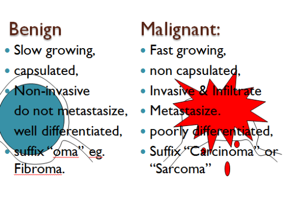
My perception of cancer was correct to some extent as cancer is caused by a tumor and tumors are masses of tissue or lumps caused by damaged cells that divide uncontrollably (MNT,2013). There are two types of tumors, benign and malignant tumors. A benign tumor is a growth that is not cancerous. This means it is localized and has not spread to other parts of the body or invaded and destroyed nearby tissue. In general, a benign tumor or condition is usually not harmful and benign tumors usually grow slowly. They can usually be removed and in most cases they never come back. However, if a benign tumor is big enough, the size and weight can press on nearby organs, blood vessels, and nerves and thus cause problems. The opposite of benign is malignant tumor. Malignant tumors are cancer, where the cancer cells can invade and damage tissues and organs near the tumor. Also, cancer cells can break away from a malignant tumor and enter the lymphatic system or the bloodstream (metastasise). This is how cancer spreads from the original tumor to form new tumors in other parts of the body (metastasise). Below is a picture showing the differences between benign and malignant tumors.

Image

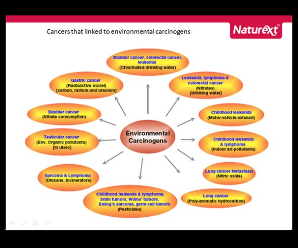
Each individual cancer is caused by a combination of things, some of which we can influence, such as lifestyle choices like smoking, and others that we cannot control, such as age and our genetics. Carcinogens which are cancer causing substances may damage cells and cause them to become cancerous due to gradual changes resulting from damage done to several of hundreds of genes that control cell growth, division and life span. If cancer were to be formed as a result of any of the above factors, the rate of growth may be slow if cancer is benign and rapid if malignant. Below is a picture that describes cancers thats linked to environmental carcinogens.

Image

When we look at cancer defined as the abnormal cell growth of a damaged cell its referred to as a relative easy concept to grasp but when we observe the clinical, cytological, and histological differences of its manifestation it becomes highly complex and therefore difficult to treat. Let I remind you that cancer is not a disease where a microorganism is identified and then a treatment method is found. Cancer is an overgrowth of the body’s own cells which make identification and treatment much more difficult. Let’s take breast cancer for example. Breast cancer is a local spread and takes place by direct invasion and infiltration between tissues (Rippey, 2012). The tumor attaches to the skin and underlying fascia or muscle of the breast. As a result of such attachment the treatment is very complicated. A mastectomy must be performed and thereafter radiation is often recommended (Treating breast cancer, 2013). In addition malignant cells adhere less strongly to adjacent cells and thus have a tendency to metastasise by themselves (Rippey, 2012).  Another good example is when tumors in the epithelial tissue invade the lymphatic system. When tumors are found in the lymph system local treatments become irrelevant and alternate treatments such as chemotherapy and the removal of the lymph come into play. The removal of the lymph would mean that the body would be left without any means to drain excess fluid in that particular area and could lead to lymph associated diseases such as lymphodema. Thus, treatment becomes very complex.Below is a video that shows how malignant tumors can spread to different areas of body.

If you have an autoimmune disease, your own immune system attacks your body tissues. Normally, our immune system protects our body against infections caused by bacteria, viruses and other parasites. But if you have an autoimmune disease, your immune cells start to attack your own normal body cells. Therefore I would say that cancer and autoimmune diseases are similar in that they both cause normal cells to malfunction and as a result cause disease in the body. Another similarity between the two is shown when cancer develops in people who have particular autoimmune diseases. For example, people who have coeliac disease have a higher risk of non Hodgkin’s lymphoma and men who have a treatment called PUVA for psoriasis can have a higher risk of penile cancer (Cancer Research UK, 2012). 

In conclusion, after reading through these modules my knowledge and understanding of cancer and autoimmunity has deepened and my perception on it has changed immensely. It is now clear to me that cancer is caused by malignant tumors and that cancers are highly complex and difficult to treat. As a result, I have great respect for patients that undergo treatments and bare the physical struggles of cancer.


References

Rippey, F F. (2012) ‘General Pathology’, Johannesburg, Witwatersrand University Press.

Martini and Nath. (2009) ‘Anatomy & Physiology’, San Francisco, Pearson Education.

Breast cancer. 2013 ‘Treatment of invasive breast cancer’ [online] Available at http://www.cancer.org/cancer/breastcancer/detailedguide/breast-cancer-treating-by-stage  [Accessed 22 April 2013]

Cancer Research UK. 2013  ’Autoimmune diseases and Cancer’ [Online] Available at: http://www.cancerresearchuk.org/cancer-help/about-cancer/cancer-questions/autoimmune-disease-and-canc [Accessed 22 April 2013]

News Medical, 2013. What is cancer. [online] Available at: www.news-medical.net/health/What-is-cancer.aspx [Accessed 22 April 2013]



Inflammation,Infection and Repair

This blog is about both acute and chronic types inflammation as well as healing and repair and infections. Inflammation is a process that is of vital importance to human health and human disease. Inflammation can be resolved through the primary reaction of acute inflammation and result in human health or can develop into chronic inflammation and result in human disease.

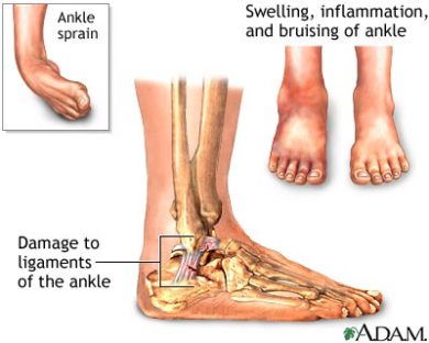
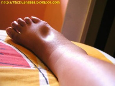
Inflammation is the body’s normal protective response to an injury,irritation, or surgery. Acute inflammation is normally a favourable response despite that the result can be uncomfortable or painful. Every person walking this earth today experienced some sort of inflammation during their life time whether it was minor or severe. For every injury that our bodies experience inflammation follows, it is the initial response of the body. Being an active person and a rugby player I tend to pick up knocks and injuries quite frequently, the most common injury of all has to be a sprained ankle. I have had a problem with my ankle since the age of 18 years and every time the injury healed I tend to injure it again within every new season. However, after the injury occurred I noticed that it displayed the cardinal signs of inflammation, there was swelling(Tumor) associated with great pain(Dolor) and redness(Rubor) as well as heat(Calor) as the swelling goes down when ice is pressed upon it and was very painful to walk on(functio laesa). It takes about two weeks if not less for the twisted ankle to heal fully provided that the ankle is left stress free. This is an example of acute inflammation. The redness and heat is due to the increased blood supply with vascular dilation which is the first bodily response. It is also useful to note that the heat generation that results from hyperaemia has an added effect of killing some foreign pathogens as these pathogens cannot survive under this warmer temperature (Biology Online, 2011). The swelling is due to oedema caused by the exudation of fluid from vessels into the tissues. The fluid that causes the oedema contains antibodies which are are used to eradicate foreign pathogens that may have entered and the increase of fluid into extracellular spaces. The pain may is due to the oedema which causes tissue tension and pressure on nerve endings. The release of chemical mediators may also cause pain. The loss of function may occur following the swelling and pain or following inflammatory destruction of tissue (Rippery, 1994). Below is a picture  of a sprained ankle that displays all the cardinal signs.

Image


After acute inflammation occurs a few things may happen. This involves healing and repair, suppuration and chronic inflammation. The one that occurs is dependent on how the tissue is damaged, the cause of the damage and the type of tissue at the site of injury(Rippery, 1994).

Healing and repair involves three stages where the damaged cells are replaced by living ones. The first stage is resolution where complete structural and functional repair occurs, the second stage would be regeneration, where restoration of parenchymal cells with some loss of normal tissue structure and lastly organisation where loss of tissue replaced with granulation and fibrous tissue resulting in the formation of a scar. Heres a video that better explains the process of healing and repair.

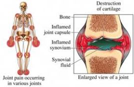
So far I have been using my ankle injury as an example of acute inflammation. I will use this example further by looking into classification of inflammation. The classification of inflammation includes the nature of injury, the site of injury and the duration of the inflammation. I would classify the inflammation I experience each rugby season as acute in duration with mild serous exudation as a sprained ankle is a moderate condition. However if a sprained ankle is left untreated or is further damaged it could cause tendon injuries which include tendonitis or swelling around the tendons. Therefore acute inflammation may go on to develop into chronic inflammation. Chronic inflammation is ‘a long lasting inflammation (weeks or months) due to persistent aggressive stimuli and is characterized by: active inflammation with mononuclear cells, tissue destruction and repair. It can follow acute inflammation or can be chronic right from the beginning’ (Danciu, 2012). A good example of chronic inflammation would be rheumatoid arthritis, where the immune system attacks the lining of the membranes that surround your joints. The resulting inflammation thickens the synovium, which can eventually invade and destroy the cartilage and bone within the joint. The tendons and ligaments that hold the joint together weaken and stretch. Gradually, the joint loses its shape and alignment. Below is a picture that further demonstrates the condition.

Image

In conclusion, I am amazed once again on our bodies ability to achieve homeostasis. Inflammation is the bodies first step to heal and thus restores human health. If inflammation is left untreated or ignored disease will occur. Therefore, the mechanism and cells involved in inflammation were made to increase health and reinstate homeostasis to the body.

References

Rippey, F F. (2012) ‘General Pathology’, Johannesburg, Witwatersrand University Press.

Martini and Nath. (2009) ‘Anatomy & Physiology’, San Francisco, Pearson Education.

Biology Online, 2013. Hyperaemia. [online] Available at: www.biology-online.org [Accessed 08 April 2013]

News Medical, 2013. What is Inflammation. [online] Available at: www.news-medical.net/health/What-is-inflammation.aspx [Accessed 08 April 2013]

 Women to women, 2011. Inflammation. [online] Available at: www.womentowomen.com [Accessed 08 April 2013]

Normal and Abnormal Fluid Distribution

Fluid and electrolyte balance plays a vital role in maintaining homeostasis in your body. The main fluid in our body is water and makes up 60% of body weight.  In this fluid there are minerals called electrolytes that are contained within the body. These electrolytes are what cells use to maintain voltages across their cell membranes and to carry electrical impulses across themselves and to other cells. Kidneys work to keep the electrolyte concentrations in blood constant despite changes in your body. Fluid intake is controlled by thirst and lost via urine, breathing, through the skin and gut. When fluid and electrolyte balance is impaired or disturbed disease will occur. These disturbances will lead to diseases such as oedema, hyperaemia and congestion, haemorrhage, shock, thrombosis and embolism and ischaemia and infarction.

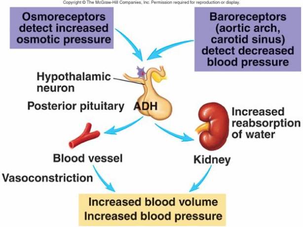
There are various hormones and mechanisms within the body that maintain fluid balance such as antidiuretic hormone (ADH) hormonal control, glomerular filtration rate, the renin-angiotensin aldosterone mechanism and through atrial natriuretic peptide release in response to atrial dilation. An example of how the body regulates fluid balance can be seen in dehydration or extreme thirst. A person is stranded in the dessert with no water, in order for the body to accommodate the loss of fluid from the extracellular fluid(ECF), the body transfers intracellular fluid(ICF) to the  ECF. Antidiuretic hormone (ADH) will also be released to increase the reabsorption of water by the kidneys. Below is a picture that further explains ADH secretion.

Image

During oedema there is the pressure within the blood vessels that pushes fluid out into the extracellular fluid. This causes increased fluid retention and ultimately swelling. It is difficult for the body to overcome oedema and treatment could include the draining of fluid from the body. When my sister was pregnant I noticed that her feet was extremely swollen and when I pressed with my finger on the enlarged area there would be a depression and would take a minute or two to return to its normal shape. It is now that I realize that she had pitting oedema. The swelling occurred because of the excess body fluid created by the baby, however, within a few days after delivery the swelling disappeared. Below is a picture that closely resembles the image that I have in my head of my sisters pitting oedema.

Image

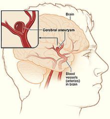
Hyperemia refers to a condition of increased blood flow to a part of the body due to dilation of arteries and arterioles. Congestion refers to a condition where there is reduce venous drainage. Having mentioned these two conditions I am reminded of a very sad incident that occurred two weeks ago. My friends brother was diagnosed with cerebral aneurysm and has been in Acoma for a couple of days. An aneurysm occurs when part of a blood vessel  or cardiac chamber swells, either the blood vessel is damaged or there is a weakness in the wall of the blood vessel. Blood pressure then builds up and inflate out at its weakest point.  According to the doctors the swelling is large and extends along  the blood vessels. The cerebral aneurism burst and caused bleeding in the brain and a hemorrhagic stroke . The doctors are currently trying to drain the blood from his brain. I hope and pray he has a successful operation. Below is a picture showing how his condition occurred.

Image

A good and very intersesting example of how the body strives to survive is in haemorrhage. Haemorrhage is when blood escapes from the blood vessels within the circulatory system and implies that there has been damage to the vessel somehow. In the case of when haemarrhage is caused by trauma by means of a motor bike accident and the person is losing blood rapidly. The persons will initially faint with dilation of the arteries to the muscles, the excessive blood loss will result in loss of consciousness. This loss of consciousness actually helps improve blood flow to the brain and the person will regain consciousness. The heart rate also increases, renin is released, plateletcount can increase causing coagulation and selective peripheral vasoconstriction occurs. These are the normal mechanisms the body undergoes to compensate for blood loss. If the blood loss of the biker is too severe he will go into Hypovolaemic shock.

Thrombosis can be seen as a disease that reacts to a problem and does further damage. Thrombosis  is the formation of a blood clot (thrombus) inside a blood vessel, obstructing the flow of blood through the circulatory system. When a blood vessel is injured, the body uses platelets and fibrin to form a blood clot, because the first step in repairing it (hemostasis) is to prevent loss of blood. If that mechanism causes too much clotting, and the clot breaks free, an embolus is formed. The formation of a thrombus is usually caused by Virchow’s triad. To elaborate, the pathogenesis includes: an injury to the vessel’s wall; by the slowing or stagnation of blood flow past the point of injury which may occur after long periods of sedentary behavior,for example, sitting on a long airplane flight; by a blood state of hypercoagulability, caused for example, by genetic deficiencies or autoimmune disorders.

In conclusion, it is amazing how our bodies react to threats and how mechanisms are in place to counter act those threats to maintain homeostasis. The process of homoestasis in regards to fluid balance is very intricate and the body has many processes to help protect and maintain the balance if it is disturbed. By reading through all these chapters I come to appreciate my body and all the mechanisms that come along with it as our bodies are truly machines that strive to survive.


References

Rippey, F F. (2012) ‘General Pathology’, Johannesburg, Witwatersrand University Press.

Martini and Nath. (2009) ‘Anatomy & Physiology’, San Francisco, Pearson Education.

Medicineplus, 2013. Fluid and Electrolyte Imbalance. [online] Available at: www.nlm.nih.gov [Accessed 24 March 2013]

News Medical, 2013. What is Thrombosis. [online] Available at: www.news-medical.net/health/What-is-Thrombosis.aspx [Accessed 24 March 2013]

Abnormal Deposition Of Material In Tissues

Amyloid

Amyloid consists of abnormal protein material. They arise from at least 18 inappropriately folded versions of proteins and polypeptides present in the body. These misfolded structures of proteins  alter its shape such that they begin to react with other cell components and form fibrils. When these Amyloid fibrils accumulate in organs, Amyloidosis occurs.

There are 5 staining methods used to recognize Amyloid. The 1st method is when iodine stains mahogany brown added with 1% sulphuric acid, which will cause the amyloid to turn dark blue. The 2nd method is Methyl Violet which stains amyloid rose pink. This is also known as a metachromatic reaction. The 3rd one is known as Congo Red and stains amyloid salmon pink. The 4th method is known as Thioflavine T which uses fluorescent dye with affinity to amyloid. The 5th and last one is known as Immunohistochemical stain and uses immunofluorescence.

Amyloid can be differentiated according to the cause of disease, whether generalised throughout the body or localised to one site, according to the chemical composition of the amyloid material deposited and the sites of deposition of the amyloid. Amyloid  can be diagnosed by histopathological means and is dependant on the staining characteristics of amyloid.

Calcification

Calcification is a gradual accumulation of calcium in an area of your body tissue. Most of the calcium absorbed by your body ends up in your bones and teeth, where it is most needed. When calcium is deposited elsewhere it is called heterotopic calcification. Excess calcium is usually dissolved in the bloodstream for excretion in the urine, but it is normal for a certain amount to collect in an area of the body tissues; this collection of calcium then hardens the tissues. This is known as dystrophic calcification. Below is an example of how calcium enters the tissues.

Image

Calcifications can be caused by inflammation or elevated levels of blood calcium, called hypercalcemia. Hypercalcemia is a cause of metastatic calcification which occurs when calcium metabolism is disturbed.  Calcification can be part of a normal healing response to musculoskeletal injuries.  Calcifications are often found in arteries affected by arteriosclerosis in benign and malignant breast processes, at sites of bone or cartilage injury, and sometimes within cancers.

Pigmentation

Pigmentation consists of 2 types of pigments, exogenous pigments and endogenous pigments.

Exogenous pigments are those which are introduced from outside the body, for example by injection, implantation, inhalation or ingestion. These pigments include dust which can be inhaled carbon particles, heavy metals that can be ingested, and miscellaneous pigments which are coloured  soluble substances derived from food.

Endogenous pigments are produced within the body and include melanin, lipofuscins and pigments formed from blood breakdown. Dark-skinned people have more melanin in their skin than light-skinned people have. Melanin is produced by cells called melanocytes. It provides some protection against skin damage from the sun. Increased melanin pigmentation may be seen in Addisons disease, in pregnancy, haemochromatosis, and in patchy or localised melanosis. Lipofuscin is a brownish pigment left over from the breakdown and absorption of damaged blood cells. Lipofuscin is found in heart muscle and smooth muscles and is also called the aging pigment. Haemosiderin is the form in which excess iron is stored in the cells and appears as golden brown pigments. Haematodin and bilirubin is the same chemically but, haematodin is a bright yellow crystaline form which may be formed locally in tissues around areas of haemarhage. Bilirubin is the pigment found in bile and gives rise to jaundice when bilirubin is in excess.

Jaundice

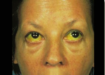
Jaundice occurs when bilirubin accumulates in the tissues and interstitial fluids of the body. Bilirubin is a yellow coloured substance that is responsible for the yellowing of the skin and sclerae. Bilirubin is a waste product that remains in the bloodstream after the iron is removed from the hemoglobin, which is released from the degradation of erythrocytes. When there is an excess of bilirubin it may leak out into surrounding tissues, saturating them with this yellow substance. In the picture below you can see this yellow substance present in the eyes and skin.

Image

Bilirubin that is circulating freely in the blood is called unconjugated bilirubin. One of the liver’s functions is to filter out waste, such as bilirubin, from the blood. Once it is in the liver, other chemicals latch on to the bilirubin, creating a substance called conjugated bilirubin, which is secreted in bile and then excreted. some of the conjugated bilirubin remains in the large intestine and is metabolized into urobilinogen, then sterobilinogen, which gives feces its brown colour.

There are a number of conditions that may cause jaundice: Acute inflammation of the liver, Inflammation of the bile duct, Obstruction of the bile duct, Haemolytic, Gilbert’s syndrome, Cholestasis, Crigler-Najjar syndrome, Dubin-Johnson syndrome, and Pseudojaundice.


References

Rippery, F.F., 1994. General Pathology. Revised edition. Witwaterstrand: Witwatersrand University Press.

Mayo Clinic, 2011. Amyloidosis. [online] Available at www.mayoclinic.com [Accessed 2 March 2013]

Medicineplus, 2013. Skin Pigmentation Disorders. [online] Available at: www.nlm.nih.gov [Accessed 2 March 2013]

Better Medicine, 2011. Calcification. [online] Available at: www.localhealth.com [Accessed 2 March 2013]

The Cell

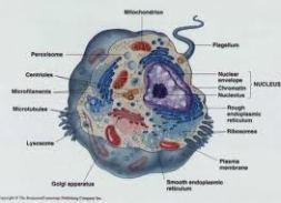
Cells are the basic units of the human body. There are many different types of cells and nearly all types of cells have the same internal structure.

There are many different types of cells of all degrees of specialisation but they all have certain features in common. The cells that are vital in the study of pathology are epithelial cells and connective tissue cells. Epithelial cells primary functions are to cover surfaces and form secretions. These cells are found on the skin, respiratory tract, alimentary tract and can be arranged to form glands of varying degrees of complexity. Connective tissue cells form another major group and include many different types of cells. Types of connective tissue cells vary enormously according to the extracellular matrix they produce. These cells can be found on tendons, bone and cartilage.

Most cells have a similar basic structure consisting of a cell membrane which surrounds the cytoplasm. The cytoplasm is the viscous content, including proteins, cell organelles, metabolites and ions. Cell organelles are membrane-bound structures such as mitochondria, the endoplasmic reticulum, the Golgi apparatus, and lysosomes. The nucleus has an envelope and contains nucleoplasm, deoxyribonucleic acid (DNA) organized as chromosomes, and the nucleolus, where ribosomes are constructed. Other structures in the cytoplasm are proteasomes, centrioles, ribosomes, the cytoskeleton, and various proteins and small molecules. The plasma membrane is the membrane surrounding the cytoplasm, and consists of a phospholipid bilayer and associated proteins and carbohydrates.

Image

Cell injury

Cells actively control the composition of their immediate environment and intracellular surroundings within a narrow range of physiological boundaries. When the cell is under physiological stress, cells can undergo adaptation to achieve a new steady state that would be well-matched with their capability in the new environment. If the injury is too severe, the affected cells die.

The causes of cellular injury are: Hypoxia and Ischemia, Chemical agents, Physical agents, Infections, Genetic defects, Nutritional defects, and Aging. More causes of cell injury include ATP depletion and the accumulation of free radicals.

Accumulation of water, fat, and other substances may also injure the cell and lead to cell death. Accumulation of water takes place in 2 stages. In the first stage the cells swell due to the accumulation of water within them. This is called cloudy swelling and injury is still reversible. This may occur in hypoxic or other forms of injury. The second stage is when hydropic generation occurs when there is substantial water logging of the cell and the vacuoles burst. At this stage the injury could be reversed but eventually the whole cell will rupture and die. Accumulation of fat in the cell is referred to as fatty change in the cell. Fat collects in cells in small membrane bound inclusions, the fat vacuoles grow in size and become visible depending on the amount of fat in the vacuole. Fatty change is more important than cloudy swelling, although it is reversible it can indicate more severe cell damage and lead to cell death. Accumulation of proteins is also known as hyaline  degeneration because of the appearance of hyaline material in the cytoplasm. Intracellular hyaline degeneration may denote serious damage to the cell and be a precursor of cell death.

Cell death and necrosis- Gangrene

Necrois is death due to unexpected and accidental cell damage. A number of toxic chemical or physical events can cause necrosis.

There are two mechanisms responsible for the development of necrosis. The first mechanism is when irreversible damage to the mitochondria and failure of ATP production. This is due to a failure of oxidative phosphorylation, anaerobic respiration will therefore continue as well as lactic acid production which accumulates causing the intracellular pH to drop, thus cell becomes more acidic. This lowered pH activates enzymes and causes the cell to digest proteins. The second mechanism is when damage to the cell membrane occurs with the loss of phospholipids.

There are 8 types of necrosis which are very important to know as it will help determine the aetiology of necrosis. The 8 types of necrosis are: Cogulative necrosis, Liquefactive necrosis or Colliquative necrosis, suppurative necrosis, Caseous necrosis, Fat necrosis, Fibrinoid necrosis, Gangrenous necrosis.

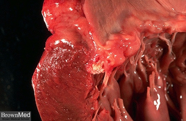
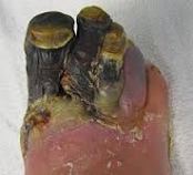
Gangrene occurs when tissue dies because its blood supply is interrupted. Gangrene may be caused by an infection, injury, or a complication of a long-term condition that restricts blood circulation. It most commonly occurs in the extremities, but internal organs and muscles may also become gangrenous. There are five main types of gangrene: Dry gangrene,Wet gangrene, Gas gangrene,  Internal gangrene, and Fournier’s gangrene.

Image


References

Rippery, F.F., 1994. General Pathology. Revised edition. Witwaterstrand: Witwatersrand University Press.

Oracle, 2013. Cellular Biology. [online] Available at: www.library.thinkquest.org [Accessed 25 February 2013]

Wiley, 2013. Cellular Injury: Mechanisms, Responses and Therapeutics. [online] Available at: www.wiley.com [Accessed 28 February 2013]

WebMD, 2012.  Skin problems and treatment: Gangrene. [online] Available at: www.webmd.com [Accessed 28 February 2013]    

Biography

I am a Chinese Medicine student at the University of the Western Cape. This course grabbed my interest because it involves treating people with the use of acupuncture and herbs. It amazes me how people can be treated by just inserting needles in to various acupuncture points on the body. It is a simple but yet very effective way of treating diseases. I am very excited to one day open my own practice.

Pathology is a very important subject because it shows you exactly what goes on in the human body. It teaches you the pathogenesis of different types of diseases that the body can encounter as well as how to recognize the symptoms of a particular disease. This module is of vital importance to me as it has all the components that will help me become a good doctor.

I have been playing sports since the age of 5years old. I played soccer from the age of 5 till 9 and then changed over to rugby and never stopped to the day. Rugby has had a positive effect on my life teaching me all kinds of qualities that is beneficial to me in everyday life. Over the years I have learnt and seen the beauty of the game and became quite good at playing it. In addition I attend the gym and rugby practice sessions at least 3 times a week to keep up with the physical demands of the game. I can therefore conclude that I am a very active person. I also enjoy watching TV, playing video games and spending good quality time with friends and family.